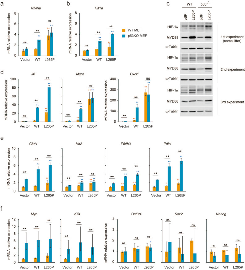
Figure 7.
Deficiency of p53 promotes increased HIF-1 signalling. (a,b,d–f) The indicated MYD88 constructs were introduced by retroviral infection to wild type and p53−/−MEFs. Expressions of the indicated mRNAs were measured by qPCR. The y-axis values are relative fold change for gene transcripts normalised to β-actin. The data represent the mean ± s.d. (n = 3) using one-way ANOVA followed by Scheffe’s F test. *P < 0.05, **P < 0.01. The black asterisks show significance between WT and p53−/−MEFs. The orange asterisks show significance between indicated cells and vector introduced WT MEFs. The blue asterisks show significance between indicated cells and vector introduced p53−/−MEFs. (c) WT and p53−/−MEFs from the same litter or same background were used for this experiment. Total cell lysates were analysed by immunoblotting.

