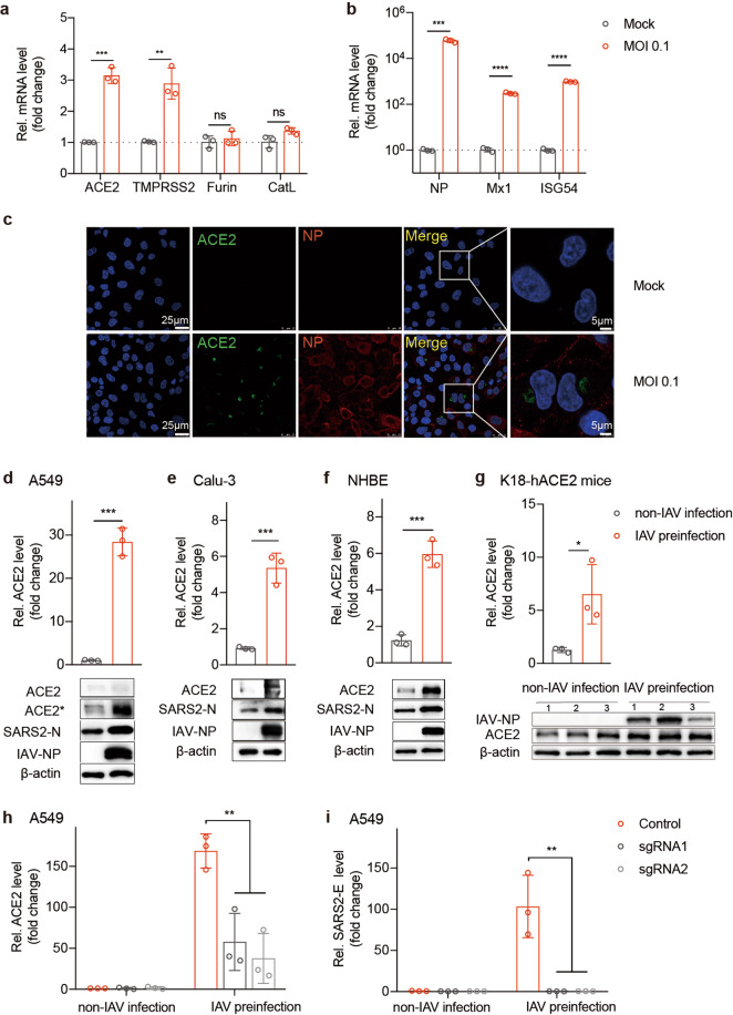
Fig. 4. ACE2 is essential for IAV promotion of SARS-CoV-2 infection.
a, b A549 cells were mock-infected or infected with WSN at an MOI of 0.1. At 12 h.p.i., total RNA was extracted from cells, and ACE2, TMPRSS2, Furin, and CatL mRNA levels (a) or NP, Mx1, and ISG54 mRNA levels (b) were evaluated via qRT-PCR using the SYBR green method. The data are expressed as fold changes relative to the mock infections. c A549 cells were infected with WSN at an MOI of 0.1. IAV NP protein (red) and ACE2 (green) were detected with an immunofluorescence assay at 12 h.p.i. Scale bars are shown. A549 (d), Calu-3 (e), and NHBE (f) cells were preinfected with WSN at an MOI of 0.1 for 12 h. Cells were then infected with live SARS-CoV-2 at an MOI of 0.01 for another 48 h. Total RNA was extracted from cells, and ACE2 mRNA was evaluated via qRT-PCR using the SYBR green method. The protein expression levels of ACE2, SARS-CoV-2 N gene, IAV NP, and β-actin were measured via western blotting assay. * means increased exposure to visualize ACE2. (g) The relative mRNA levels of ACE2 were measured in lung homogenates from the indicated groups, and the protein expression of IAV NP and ACE2 was detected via western blotting. (d–g) The data are expressed as fold changes relative to the non-IAV infection control. (h–i) To establish ACE2 knockdown cells, A549 cells were transduced with lentivirus encoding the CRISPR-Cas9 system with two guide RNAs targeting ACE2 (sgRNA1 and sgRNA2) or control guide RNA. Cells were infected with live SARS-CoV-2 at an MOI of 0.01 with or without IAV infection using the same procedure described above. The ACE2 (qRT-PCR) (h) and SARS-CoV-2 E gene (Taqman-qRT-PCR) (i) mRNA levels were detected. The data are expressed as the fold change relative to the non-IAV infection control. Values represent means ± SD of three independent experiments. *P < 0.05, **P < 0.01, ***P < 0.001, ****P < 0.0001.

