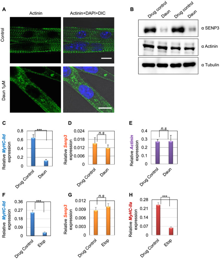
Figure 7.

Effect of drug treatment on stem cell‐derived muscle cells. (A) Satellite stem cells were isolated from wild‐type C57BL/6J adult mice (30 days old) followed by differentiation into matured myotubes. After overnight daunorubicin (Daun) treatment, immunofluorescence staining followed by confocal microscopy was performed with an antibody against alpha‐actinin to probe for Z‐disc striation of a fully matured primary myotubes in control cells as well as Daun‐treated cells. The size of both the scale bars—10 μm each. DIC, differential interference contrast microscopy. (B) Western blots show status of indicated protein expression in response to drug treatment in primary myotubes. (C–E) Expression of indicated mRNAs were monitored in quantitative reverse transcription polymerase chain reaction assay. Data represent average (± SEM) of stem cells isolated from two different mice, with technical duplicates for each experiment. Stars indicate statistically significant difference, unpaired t‐test, P = 0.0004. n.s, not significant. (D–H) Similar to (C), except the primary myotubes were treated overnight with etoposide (Etop). Data represent average (± SEM) of primary myotubes from two different mice, with technical quadruplicates for each experiment. Stars indicate statistically significant difference, unpaired t‐test, P < 0.0001. n.s, not significant.
