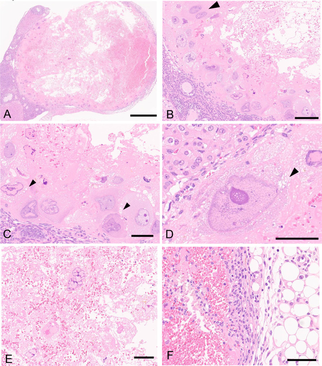
Fig. 1.
Histopathology the ovary of a young female Crl:CD1 (ICR) mouse stained with hematoxylin and eosin. (A) A cystic mass containing abundant blood plasma and erythrocytes. The mass is well defined from normal ovarian tissues and peripheral adipose tissues. Bar = 500 μm. (B) Large pleomorphic tumor cells with bizarre shaped abnormally shaped nuclei are arranged at the edges of the mass. Note: binucleated cell (arrowhead), blood plasma, and erythrocytes are observed. Bar = 100 μm. (C, D) The tumor cells possess abundant eosinophilic to amphophilic cytoplasm, which is occasionally vacuolated (arrowheads). The tumor cells contain a single large nucleus, and dark-stained chromatin is occasionally observed near the nuclear membrane. Bar = 50 μm. (E) Tumor cells located in the center of the mass are frequently degenerative or necrotic. Bar = 50 μm. (F) Inflammatory cells have infiltrated the edge of the mass and in the peripheral adipose tissue. Bar = 50 μm.

