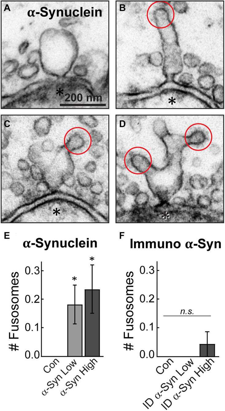
FIGURE 7.
Excess brain-derived human α-synuclein produces abnormal fusion/fission events. (A–D) Electron micrographs showing morphology of “fusosomes” in the active zone induced by excess α-synuclein. Red circles indicate CCPs emanating from some fusosomes. Asterisks indicate post-synaptic dendrites. Scale bar in A = 200 nm and applies to B–D. (E,F) Quantification reveals a significant increase in the number of fusosomes per synapse after treatment with α-synuclein (n = 34–37 synapses, two axons), but not with the immunodepleted sample (n = 21–23 synapses, two axons). Data represent mean ± SEM per synapse per section. “ID” indicates “immunodepleted.” Asterisks indicate statistical significance (p < 0.05), and “n.s.” indicates “not significant” by ANOVA.

