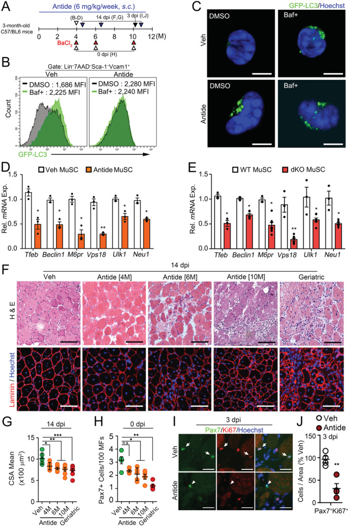
Figure 4.

Impaired autophagosome clearance and regenerative function of MuSCs under reduction of HPG‐sex steroid hormone‐Tfeb axis. (A) A scheme for Antide treatment. Three‐month‐old mice were subcutaneously injected with vehicle (Veh) or Nal‐Lys GnRH antagonist (Antide) for 4, 6, or 10 months. (B and C) MuSCs were isolated from Veh or Antide‐treated GFP‐LC3 transgenic mice for 4 months and treated with DMSO or Baf for 6 h. Flow cytometry (B) and representative confocal images (C) for GFP‐LC3 in MuSCs. (D and E) Tfeb and Tfeb target‐gene expressions in MuSCs isolated from Antide‐treated mice in Figure 4B (D) and dKO mice in Figure 1A (E). (F) TA muscles of Antide‐treated mice for indicated time were injured by BaCl2 injection and were analysed at 14 dpi. Haematoxylin and eosin (H&E) staining (top) and IHC staining for laminin (bottom). Geriatric mice were 30 months old. (G) Quantifications of CSA of regenerating MFs. (H) The number of Pax7 + cells per 100 MFs in uninjured Veh, Antide‐treated, and geriatric TA muscles. (I and J) TA muscles from Antide‐treated mice for 10 months were injured by BaCl2 and analysed at 3 dpi. Representative images for Pax7 and Ki67 staining (I) and quantifications (J). Arrows and arrowheads indicate Pax7 +Ki67 + and Pax7 +Ki67 − cells, respectively. Scales, 5 (C), 50 (I), and 100 μm (E). Comparisons by one‐way ANOVA with Tukey's post hoc test (G–H), Mann–Whitney U test (J) and unpaired t‐test (D–E). Bars, mean ± SEM; n = 4–5 animals per group; *P < 0.05, **P < 0.01. dKO, double knockout; MuSCs, muscle stem cells; WT, wild‐type.
