
Figure 6.
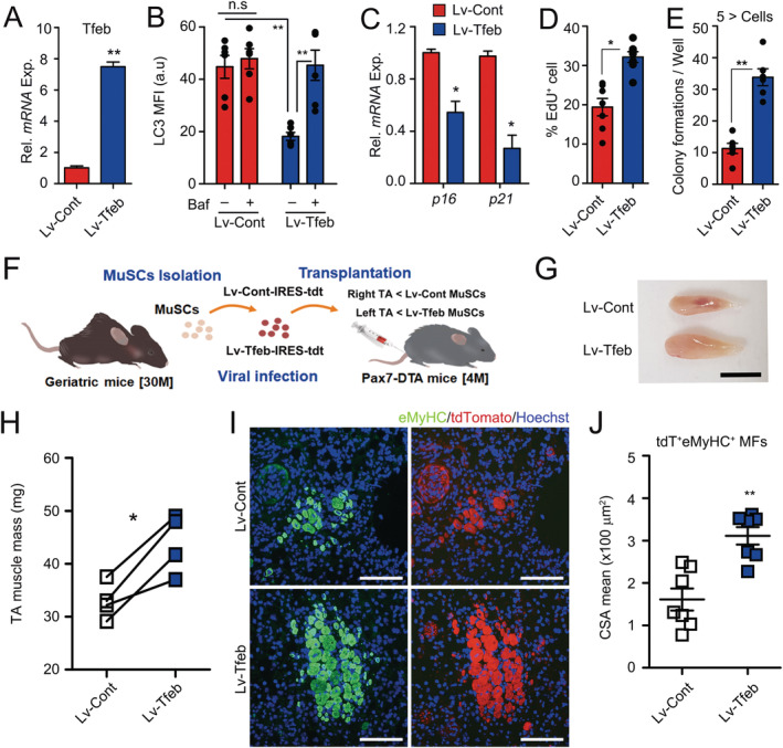
Improved function of geriatric muscle stem cells (MuSCs) by Tfeb overexpression. (A–E) Geriatric MuSCs were transduced with Lv‐Cont and Lv‐Tfeb lentivirus. Seventy‐two hours after transduction, Tfeb expression (A) and quantification of MFI for LC3 proteins with or without Baf for 6 h (B). Relative expressions of p16 and p21 in transduced MuSCs (C). Percentages of EdU + cells (D) and number of colony formation (E) 96 h after transduction. Representative images for EdU incorporation and colony formation assays of the MuSCs were shown in Figure S7D and S7E. Cell clusters consisting of at least five or more cells were counted as colony. (F) A scheme for MuSC transplantation. Equal numbers of 16 h of transduced geriatric MuSCs were transplanted into young injured Pax7CreER;ROSA‐DTA mice pre‐treated with tamoxifen for 4 weeks before injury. Ten days after MuSC transplantation, the TA muscles were analysed. (G) Gross morphology and (H) muscle mass of TA muscles. (I) IHC images for eMyHC and tdTomato, and (J) quantification of mean CSA for tdT + eMyHC + myofibres. Scales: 0.5 cm (G) and 100 μm (I). Comparisons by Mann–Whitney U test (B, D, E, and H) and unpaired t‐test (A, C, and J). Bars, mean ± SEM; n = 4–7 animals per group; *P < 0.05, **P < 0.01, n.s. not significant.