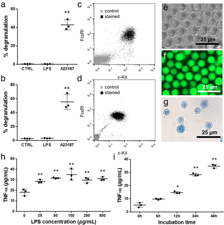
FIGURE 1.

Identification of BMMCs and PCMCs. Mast cell nature was confirmed by showing the degranulation capacity of BMMCs (a) and PCMCs (b) with β‐hexosaminidase assay in the presence of LPS (100 ng/ml ) and A23187 (0.5 μM). Cell purity was assessed by flow cytometry on the 4th week of mast cell differentiation (BMMC, c) or on 10th day of culture (PCMC, d) by flow cytometry, based on FcɛRI and c‐Kit positivity. BMMCs from C57BL/6 mice were studied by phase contrast microscopy (e), while GFP‐BMMCs from C57BL‐GFP mice were analysed by fluorescent microscopy (f). Cellular purity was also checked by Kimura staining (g). The optimal concentration of LPS (h) and incubation time (i) were chosen based on TNF‐α secretion measured by ELISA. Graphs show the mean and SD of three biological replicates, where one point indicates the average of three technical replicates (n = 3, *P ≤ 0.05, **P ≤ 0.01 t test)
