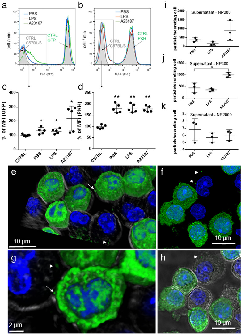
FIGURE 2.

Uptake of MC‐derived EVs by MCs. Non‐fluorescent BMMCs from C57BL/6 mice were co‐cultured with GFP‐BMMCs with cytoplasmic fluorescence, derived from C57BL‐GFP mice (a, c) or PKH‐stained BMMC with fluorescent plasma membrane (b, d) in the presence of PBS or 100 ng/ml LPS. A23187 (0.5 μM) was used as positive control. For negative control, unstained C57BL/6‐derived BMMCs were used (a and b, grey histograms). Scatter plots show the mean and SD of five biological replicates (n = 5), normalized to unstained control. Cell‐free conditioned medium of BMMCs (cultured upon addition of PBS, 100 ng/ml LPS and 0.5 μM A23187) was tested by TRPS with three different pore size membranes (i: NP200 for small EVs, j: NP400 for mEVs and k: NP2000 for lEVs). Graphs show the mean and SD of three independent experiments (n = 3), where one point indicates the average of two technical replicates. e‐h: GFP‐BMMC and wild type BMMCs were co‐cultured for 24 h. Cells were fixed with 2% PFA, nucleus was stained with DAPI and EV‐transfer from GFP‐BMMCs was tested with confocal microscopy (Leica SP8). Cell shapes were visualized by transmitted light. 3D images were generated using LAS X software. Arrows: Releasing EVs, Arrow heads: EVs
