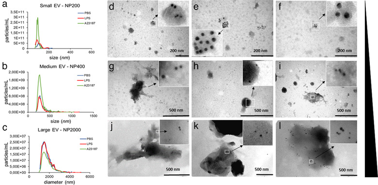
FIGURE 3.

Immune electron microscopic and TRPS analyses of MC‐derived EVs. Particle sizes, size distributions and concentrations of separated large‐, medium size‐ and small‐EVs from the conditioned medium of BMMCs were measured by TRPS using membranes with three different pore sizes (a: NP200 for sEVs, b: NP400 for mEVs and c: NP2000 for lEVs. BMMCs were cultured in the presence of PBS (negative control, blue), LPS (100 ng/ml, red) or A23187 (positive control, green) for 24 h prior to EV separation. Graphs are means of 3 biological replicates with 2‐2 technical replicates (n = 6). EV markers of BMMC‐derived small (d‐f), medium (g‐i) and large (j‐l) EVs were detected by immunoelectron microscopy using nanogold labelling. PBS (unstimulated): 1st column, LPS‐stimulated: 2nd column, and A23187‐stimulated: 3rd column. Gold particles with 10 nm diameter represent CD63 while 5 nm diameter particles indicate c‐Kit
