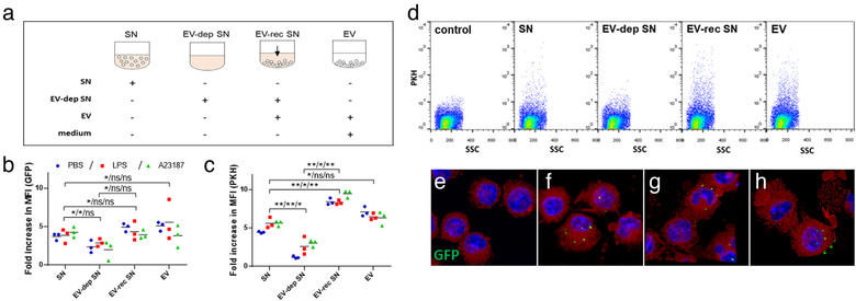
FIGURE 5.

LPS‐stimulated mast cell derived extracellular vesicles are taken up by other mast cells. GFP‐BMMCs (b) and PKH‐stained BMMCs (c, d) were cultured in EV‐free medium in the presence or absence of LPS (100 ng/ml ) for an hour. A23187 (0.5 μM) and PBS were used as positive as negative controls, respectively. EVs were separated from the conditioned medium after 24 h . Unstained BMMCs derived from C57BL/6 mice were cultured in EV‐free medium (control), conditioned medium (SN), EV‐depleted conditioned medium (EV‐dep SN), EV‐reconstructed medium (EV‐rec SN) or separated EVs in fresh medium (a). EV‐uptake was also monitored by confocal microscopy (e‐h: GFP (green) merged with DAPI (blue) and lactadherin (red), e: control BMMCs; f: BMMCs cultured with EVs derived from PBS‐stimulated, g: LPS‐stimulated and h: A23187‐stimulated MCs. The mean of flow cytometric fold increase in MFI values (b, c) normalized to unstained control of three biological replicates (n = 3, *P ≤ 0.05, **P ≤ 0.01 t test) are shown. Flow cytometric plots (d) show single demonstrates of three independent experiments
