FIGURE 6.

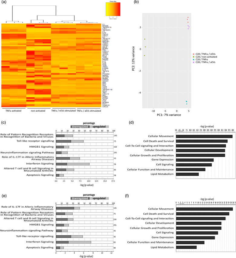
M‐EVs are multimodal signaling mediators in TNFα‐activated cellular inflammation. (a) Hierarchical clusterization of 50 differentially expressed genes in C20 microglia stimulated with a combination of TNFα and nEVs or aEVs compared to only TNFα−activated control cells and non‐activated control cells. Yellow marks low expression and orange‐red marks high expression (n = 3 per group). A complete list of differentially expressed genes can be found in Tables ST3‐ST4. (b) PCA plot showing disparities of microglia activated with TNFα, either non‐stimulated or M‐EVs stimulated. (c,d) List of top significant IPA canonical pathways and molecular function and their P ‐values in TNFα‐activated microglia versus TNFα/nEVs stimulated microglia and (E,F) TNFα/aEVs stimulated cells compared to TNFα‐activated control cells. All RNA sequencing data represent the average of three biological experiments
