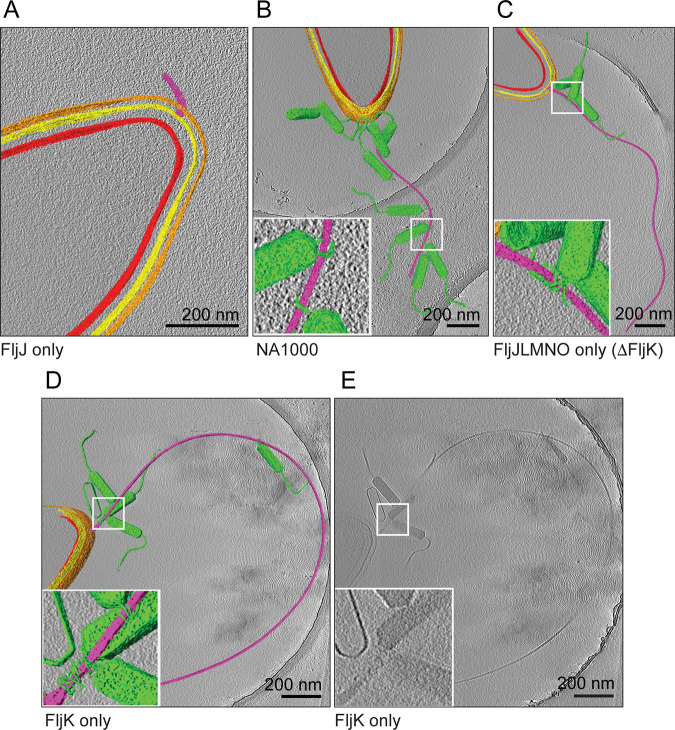
FIG 4.
Averaged slices merged with segmentations through three-dimensional tomographic reconstructions of representative ϕCbK-infected C. crescentus cells, selected from all strains examined. Segmentation was performed in Amira, coloring the inner membrane red, outer membrane yellow, S-layer orange, flagellum purple, and phage particles green. Scale bars are 200 nm. (A) C. crescentus FljJ assembles only a shortened flagellum. (B, C, and D) Phages were observed either adsorbed along the length of the flagellum or attached to the cell poles of strains investigated. Insets highlight the ϕCbK head filament wrapped around the C. crescentus flagellum. (E) Unsegmented tomographic slice of FljK for comparison.

