FIGURE 5.

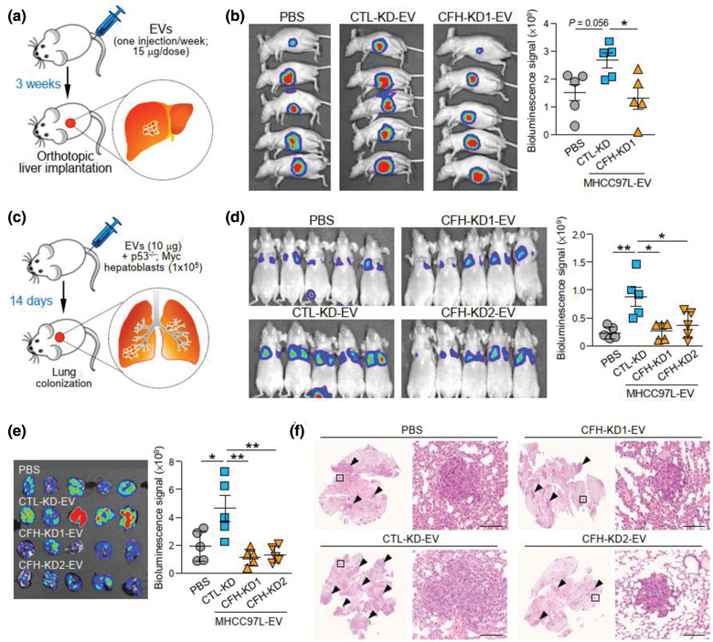
EV‐CFH promotes HCC tumorigenesis and metastasis. (a) Schematic diagram of EV education model. EVs derived from MHCC97L control (CTL‐KD) and CFH knockdown (CFH‐KD1) cells were injected into mice through tail vein once a week for 3 weeks (15 μg per week) prior to orthotopic liver implantation of luciferase‐labelled MHCC97L tumour seed (n = 5). Development of liver tumour was analysed 6 weeks after liver implantation. (b) Bioluminescence imaging of animals at the end of experiment. Quantification of luciferase signal is plotted. (c) Diagram illustrating the procedure of experimental metastasis assay. Murine p53‐/‐;Myc hepatoblasts were injected intravenously with or without EVs of MHCC97L control (CTL‐KD) or CFH knockdown (CFH‐KD1) cells (n = 5). Animals were subjected to bioluminescence imaging 14 days after injection. (d) Bioluminescence imaging of animals at the end of experiment. Intensity of luciferase signal is quantified. (e) Ex vivo bioluminescence imaging of dissected lung tissues. Quantification of luciferase signal is shown. (f) Representative images of H&E staining of lung tissues. Examples of metastatic lesions are indicated by arrowheads. Insets indicate the enlarged area of the metastatic lesions. Data are represented as mean ± SEM. * P < 0.05, ** P < 0.01. P < 0.05 is considered as statistically significant
