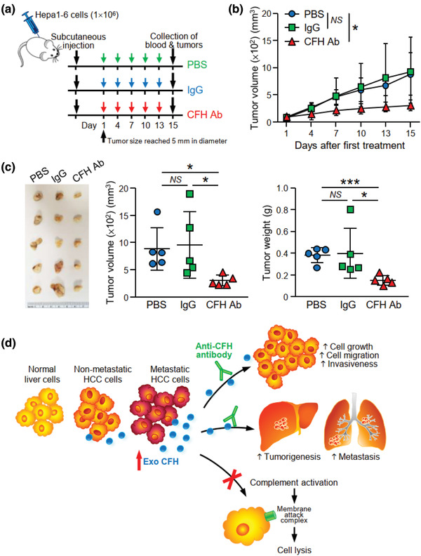
FIGURE 8.

Anti‐CFH antibody treatment suppresses tumour development in syngeneic mouse model. (a) Schedule of anti‐CFH antibody administration in mice. Hepa1‐6 cells were injected subcutaneously into C57BL/6N mice. Administration of antibody was started when the tumour size reached 5 mm in diameter. PBS, control IgG or murine anti‐CFH antibody (200 μg/mouse) were administered by intraperitoneal injection once every 3 days for 2 weeks. (b) Tumour size was measured regularly during the experimental period and plotted. (c) Tumours developed were excised and tumour size and weight were measured. Image showing the fixed tumours (left). Tumour volume (middle) and weight (right) were plotted. (d) A proposed model illustrating the role of EV‐CFH in HCC. EVs with higher CFH level are derived from HCC cells with higher metastatic potential when compared to EVs of non‐metastatic HCC cells and normal liver cells. EV‐CFH promotes HCC cell growth and motility in culture and tumorigenesis and metastasis in animals. Uptake of EVs also protects HCC cells from complement‐mediated cytotoxicity, thus facilitating the survival and growth of cells in liver and distant lung tissues. Data are represented as mean ± SEM. * P < 0.05, *** P < 0.001. P < 0.05 is considered as statistically significant. NS, not significant
