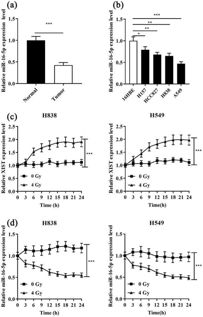
Figure 3.

Opposite changes in the expressions of XIST and miR-16-5p in NSCLC cells under irradiation. (a) and (b) qRT-PCR was used to detect the expression of miR-16-5p in four NSCLC tissue and cell lines (16HBE). (c) XIST expression in H838 and A549 cells was detected under X-ray irradiation at a dose of 4 Gy every 3 h. (d) MiR-16-5p expression in H838 and A549 cells was detected under X-ray irradiation at a dose of 4 Gy every 3 h.
*P < 0.05, **P < 0.01 and ***P < 0.001.
