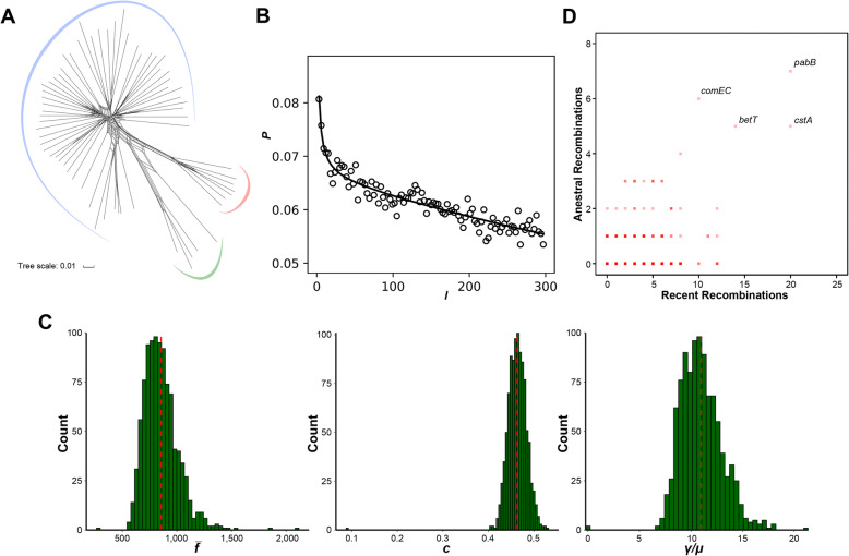
Fig. 4.
Recombination in M. luteus. a Phylogenetic network inferred with the concatenation of single-copy core genes. Lines between splits show where recombination has occurred. Three major clades are presented by different colored arcs. The scale bar indicates 1% sequence divergence. b Correlation profile (circles) calculated by mcorr. Model fit is shown as a solid line. c Distributions of the three recombination parameters for all pairs of genomes. Red vertical lines indicate the means calculated with 1000 bootstrapped replicates. d Core genes that have undergone recent and ancestral recombination. Horizontal and vertical axes show the estimated number of recent and ancestral recombinations, respectively. Names of some of the most frequently recombined genes are shown

