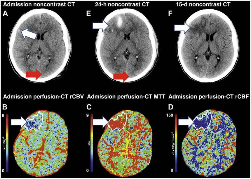
Figure 2:

Contrast-enhanced and perfusion-CT images from a patient with severe TBI. (A) Noncontrast CT at admission demonstrates a small hemorrhagic contusion in the right frontal lobe (arrow). Admission perfusion-CT images demonstrate a large territory of decreased rCBV (B), increased MTT (C) and decreased rCBF (D). Follow-up noncontrast CT at 24 hours (E) demonstrates increased areas of hemorrhagic contusion in the right frontal lobe where the perfusion abnormality was seen. Follow-up noncontrast CT at 15-days (F) demonstrates evolving hemorrhagic contusion and encephalomalacia in the right frontal lobe, which corresponds to the same distribution that is seen on the perfusion-CT.
