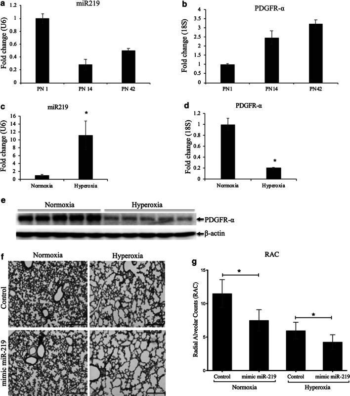
Fig. 4.
miR-219-5p (miR-219) and PDGFR-α during lung development and effect of hyperoxia and mimic miR-219 on miR-219 in a BPD mouse model. a Expression of miR-219 in lung homogenates from mice at 4, 7, and 14 days of age (P1, P14, and P42). b Expression of PDGFR-α by quantitative RT-PCR (qPCR) at P1, P14, and P42, showing increase PDGFR-α at P14, and P42 when miR-219 is decreased. Lung homogenates from mice at 14 days of age exposed to air or hyperoxia were analyzed by qPCR of RNA from lung homogenates for miR-219 (c) and PDGFR-α (d) or by western blot for PDGFR-α (e). f Representative photomicrographs of H&E-stained sections of alveolar regions from lungs of mice at 14 days of age administered either with mimic miR-219, or control mimic. g Radial alveolar counts (RAC) of mice administered control vs mimic miR-219 in normoxia and hyperoxia. Values are means ± SE; n = 6 mice/group. *P ˂ 0.05 vs. corresponding group

