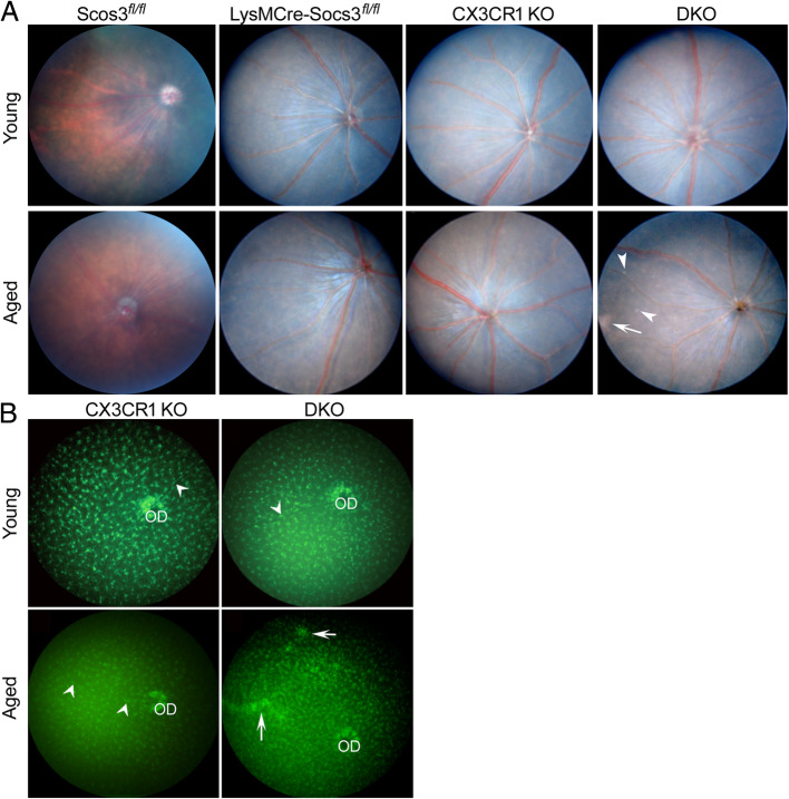
Fig. 1.
Fundus images and green fluorescence images of different mice. a Fundus images were taken using the topic endoscopic fundus imaging (TEFI) system from young (3-month) and aged (12-month old) Socs3fl/fl mice, LysMCre-Socs3fl/fl mice, Cx3cr1gfp/gfp (CX3CR1 KO) mice, and DKO mice. Arrow – a patch of whitish lesion; arrowheads – multiple whitish dots. b Fundus green fluorescence images were taken from 3-month or 12-month old Cx3cr1gfp/gfp and DKO mice using Micron IV. Arrows – patches of GFP aggregations; arrowheads – perivascular macrophages. OD – optic disc

