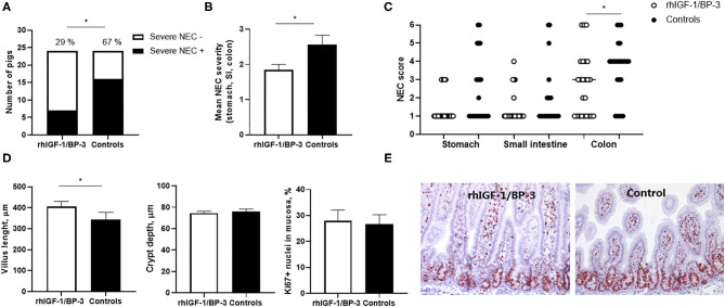
Figure 3.
Intestinal lesions and structure. (A) Incidence of severe NEC (defined as score ≥4 in at least one gut region) in rhIGF-1/BP-3 and control preterm pigs (n = 24). (B) NEC severity score across gut regions in rhIGF-1/BP-3 and control preterm pigs (n = 24). (C) Individual NEC scores in stomach, small intestine regions (highest score of the proximal, middle and distal section) and colon regions (highest score of the colon and cecum). (D) Villus length, crypt depth and Ki67 stained area relative to total nuclei area in tunica mucosa in mid small intestine (n = 23). Values are mean±SEM. (E) Immunohistochemically staining for Ki67 with hematoxylin counterstain in a representative middle small intestine section from a preterm pig treated with rhIGF-1/BP-3 and a control preterm pig. Pictures are 10x. *p < 0.05, **p < 0.01, ***p < 0.001.

