
Figure 6. Histology of the Trpm3-mutant lens.
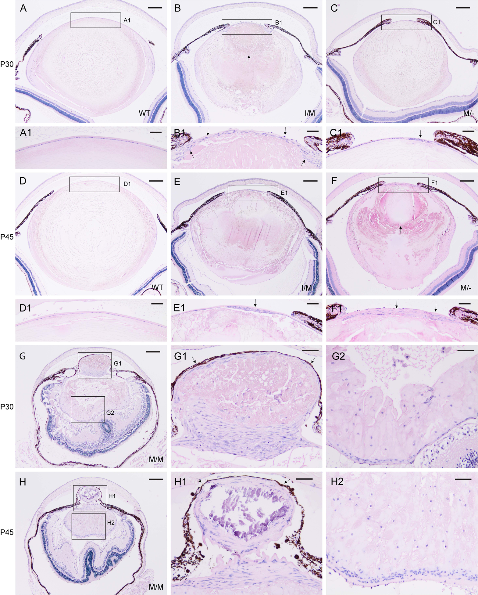
Representative H&E staining of lenses from wild-type (A, D), Trpm3-I/M (B, E), Trpm3-M/- (C, F), and Trpm3-M/M (G, H) mutant mice at P30 (A-C, G) and P45 (D-F, H) showing progressive lens degeneration and accumulation of nucleated cells (B, C, E, F, G, H, G1, G2, H1, H2). Arrows indicate abnormal multi-layering of the lens epithelium (B1, C1, E1, F1), anterior displacement of the lens nucleus (B, F), and iris or melanin pigment adhesion to the anterior lens capsule (G1, H1). In addition to cell nuclei, hematoxylin stained blue/black mineralization-like deposits (H1). Scale bar: 200 μm (A-H), 50 μm (A1–H1, G2, H2).