
Figure 7. Mineralization of the Trpm3-mutant lens.
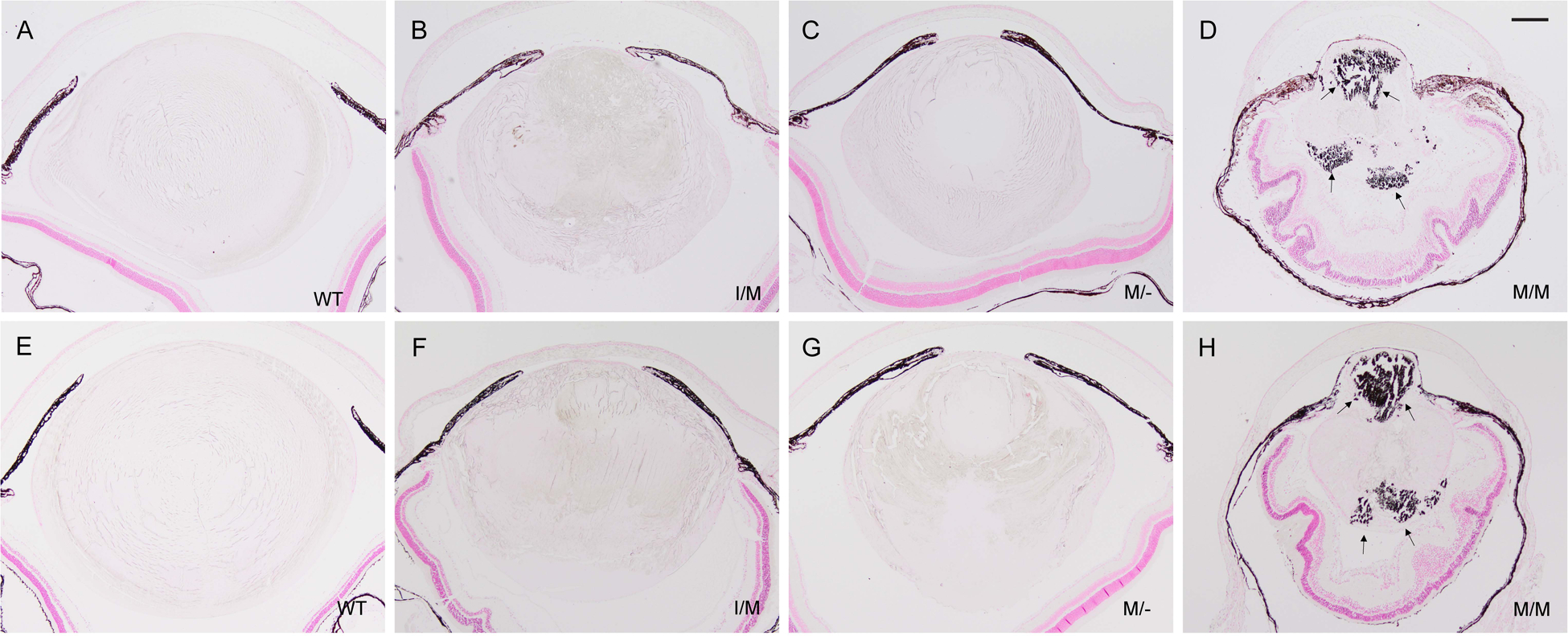
Representative von Kossa (silver) staining of lenses from wild-type (A, E), Trpm3-I/M (B, F), Trpm3-M/- (C, G), and Trpm3-M/M (D, H) mutant mice at P30 (A-D) and P45 (E-H) showing progressive accumulation of calcium phosphate-like (black) deposits (D, H arrows). A-H counterstained with nuclear fast red. Note, the iris anterior to the lens (A-H) and the retinal pigment epithelium posterior to the lens (D, H) appear dark brown/black due to melanin pigmentation. Scale bar: 200 μm.