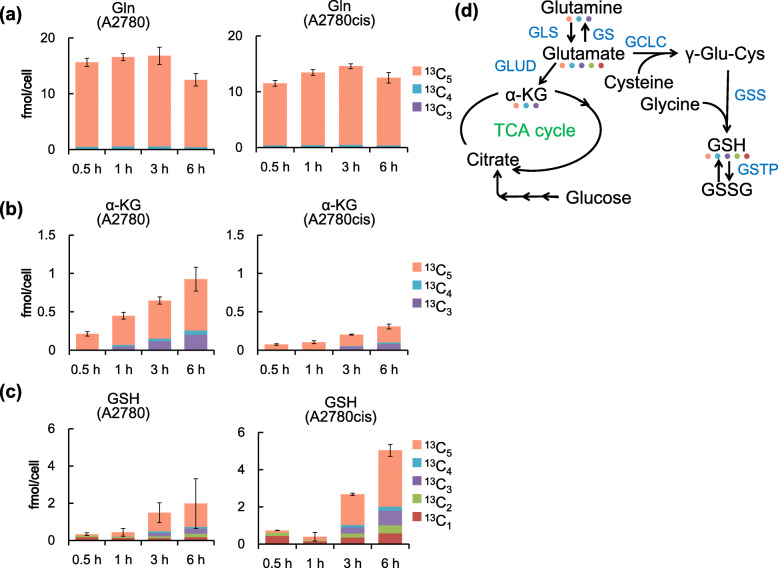
Fig. 2.
Metabolic flux analysis using isotopically labelled glutamine in A2780 and A2780cis cells. a, b, and c Isotopologue distribution of metabolites in A2780 and A2780cis cells. Cells were incubated with medium containing glutamine isotopically labeled at all five carbon atoms (13C5-glutamine) for the indicated time periods. Carbon fluxes from glutamine to Glu, GSH, and α-ketoglutarate (α-KG) were determined using CE-TOFMS. Each bar color corresponds to the number of 13C replaced with 12C in the metabolites. Data are shown as the mean ± SD of three independent experiments. d A pathway map of glutamine metabolism. Metabolites and catalytic enzymes are shown in black and blue, respectively. The colored dots show the 13C isotopically labeled metabolites, and the color corresponds to the icons in Fig. 2a–c on the left

