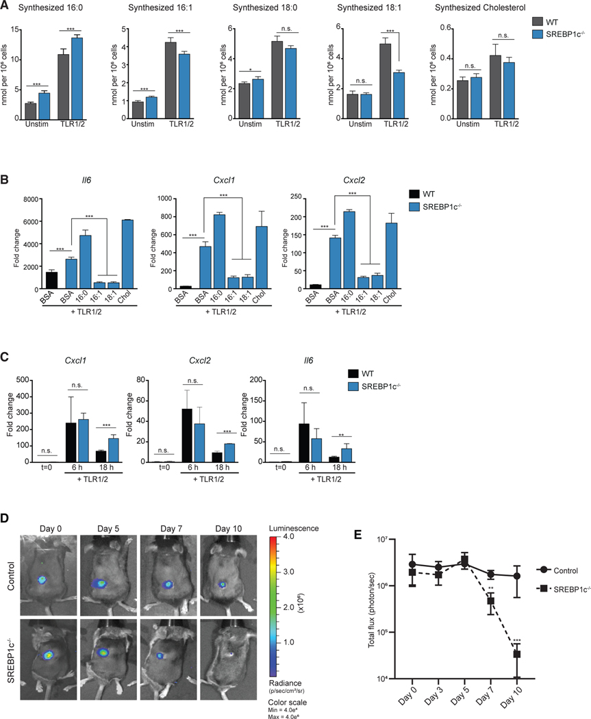
Figure 7. Upregulation of SREBP1c Is Required for MUFA Flux to Control Inflammation.
(A) Net synthesized palmitic acid (16:0), palmitoleic acid (16:1), stearic acid (18:0), oleic acid (18:1), and cholesterol from WT control or SREBP1c−/− BMDMs stimulated with TLR1/2 agonist.
(B) qPCR analysis of inflammatory gene expression from WT control or SREBP1c−/− BMDMs stimulated with TLR1/2 agonist ± BSA-conjugated 16:0, 16:1, or 18:1 fatty acids, or cholesterol for 24 h.
(C) qPCR analysis of inflammatory gene expression from cells collected by peritoneal lavage from WT control or SREBP1c−/− mice injected (intraperitoneal) with TLR1/2 agonist for 6 h and 18 h.
(D) Time course bioluminescence images from a representative WT control and SREBP1c−/− mouse from day 0 (immediately post-infection) through day 10 post-infection challenged with the bioluminescent strain of Staphylococcus aureus (Xen36).
(E) Time course quantification of total flux (photons/sec) from WT control or SREBP1c−/− mice infected with bioluminescent S. aureus.
All isotope labeling experiments are from four biologic replicates per experimental condition and representative of greater than three experiments. Gene expression studies are from three biologic replicates per experimental condition and are representative of greater than three experiments. In vivo experiments with S. aureus infections or TLR1/2 agonists are representative of four independent experiments.
All data are presented as mean ± SEM. *p < 0.05; **p < 0.01, ***p < 0.001.

