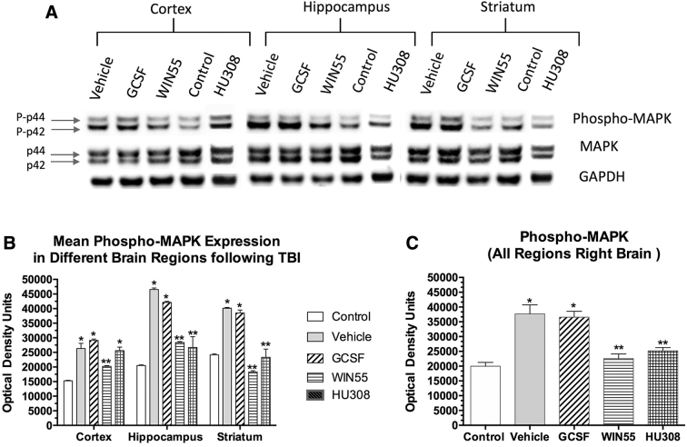
FIG. 4.
Effects of TBI followed by treatment with G-CSF and cannabinoids on signal transduction (phospho-MAPK protein expression). (A) Sample Western blot showing phospho-MAPK and GAPDH expression following various treatments. (B) Quantification (optical density) of normalized signal in three brain regions from three mice per treatment (from right hemisphere). Each bar is the mean (±SEM). Two-way ANOVA, followed by correction for multiple comparisons, revealed that both the brain region and treatment contributed significantly to total variance (p<0.0001); *p<0.001 compared with control (sham surgery, no treatment) and **p<0.01 compared with vehicle-treated samples. (C) Analysis of pooled data from the three regions of the right hemisphere (n=9; three samples from each of the three mice per treatment). MAPK, mitogen-activated protein kinase.

