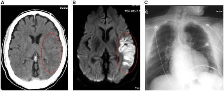
Figure 1.
Representative brain computed tomography and magnetic resonance imaging and chest X-ray of the patient. (A) Brain computed tomography demonstrating subtle changes in the left middle cranial artery territory. (B) Brain magnetic resonance imaging demonstrating left middle cranial artery infarct. (C) Chest X-ray demonstrating clear lung fields with normal cardiac silhouette.

