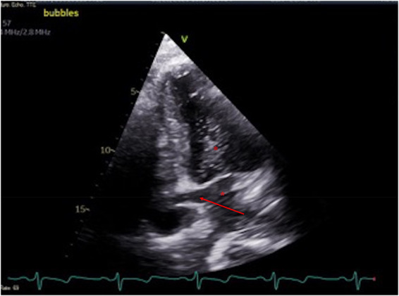
Figure 3.

Echocardiographic image of the heart demonstrating unroofed coronary sinus (arrow) and appearance of the microbubbles in the coronary sinus, left atrium, and left ventricle (asterisk) in modified apical four-chamber view (posterior angulation).
