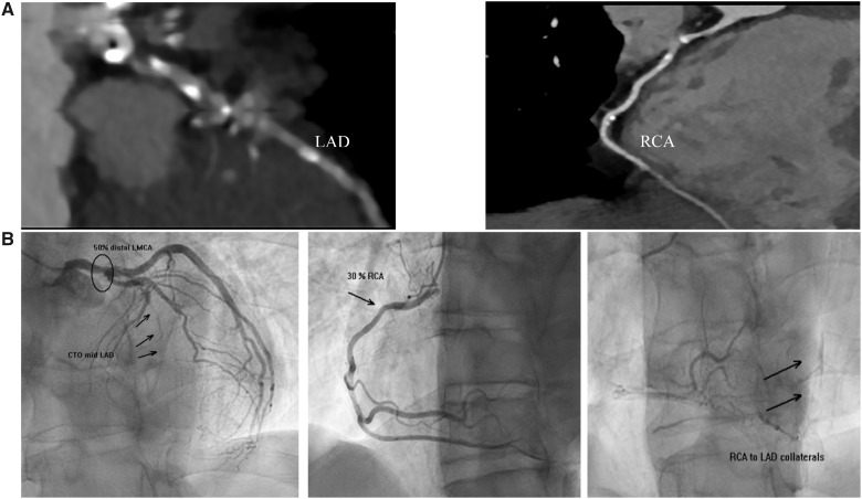
Figure 5.
Representative computed tomography coronary angiography and coronary angiogram of the patient. (A) Computed tomography coronary angiography demonstrating calcification and plaque in the left anterior descending and right coronary arteries. (B) Coronary angiography demonstrating 50% distal left main lesion, chronic total occlusion of the mid left anterior descending, and 30% right coronary artery lesion with collaterals from right coronary artery to left anterior descending. CTO, chronic total occlusion.

