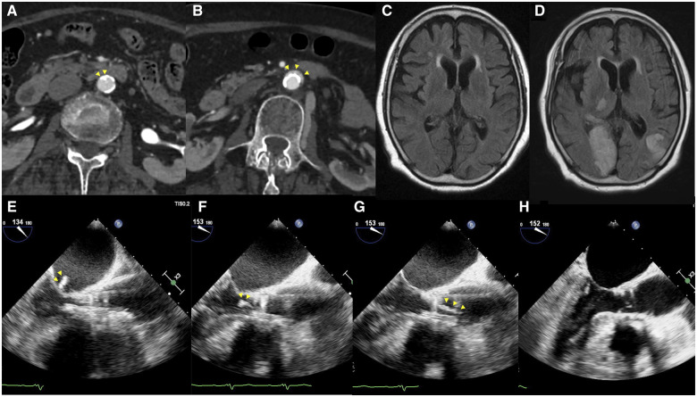
Figure 1.
(A) Abdominal computed tomography prior to transcatheter aortic valve implantation. A 5 mm calcified plaque can be seen in the lumen of the abdominal aorta (arrowhead). (B) Abdominal computed tomography after transcatheter aortic valve implantation. Localized aortic dissection can be seen (arrowhead). (C) Cerebral magnetic resonance imaging prior to transcatheter aortic valve implantation. (D) Cerebral magnetic resonance imaging after transcatheter aortic valve implantation. Acute cerebral infarction was found on magnetic resonance imaging after transcatheter aortic valve implantation. The patient presented with hemianopsia and ataxia. (E–H) Transoesophageal echocardiography during transcatheter aortic valve implantation; mid-oesophageal long-axis views. A high echoic, 10 mm long mass (arrowhead) is attached to the transcatheter heart valve (E). Subsequently, the mass is moving in and out of the left atrium and the left ventricle (F). After implantation, the mass gradually enlarged to a length of 26 mm (G). Then, the mass spontaneously detached from the transcatheter heart valve and flowed up the ascending aorta, disappearing from the transoesophageal echocardiography field of view (H).

