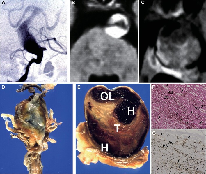
FIGURE 11.
A mortality case with conservative management. A 66-yr-old man presented with “locked-in” syndrome caused by a large fusiform aneurysm of the basilar trunk. A, Right vertebral artery angiogram demonstrating dolichoectatic basilar artery. Bilateral anterior inferior cerebellar arteries are coming off from this lesion. B, Magnetic resonance angiogram (MRA) demonstrated a tortuous, enlarged basilar artery with a diameter of 1.5 cm with intramural hemorrhage within the aneurysm wall. C, Follow-up MR image 4 yr later revealed progressive aneurysm growth exceeding 4 cm. D, Five years later, he suddenly developed hypotensive shock and deceased. Postmortem examination revealed a giant aneurysm arising from the middle one-third of the basilar artery. E, Coronal section of the aneurysm showed an open lumen (OL), flap-like tissue (arrow) containing numerous vascular channels, staged laminated thrombus (T), and thick aneurysm wall. The staged thrombus contains hemorrhage (H) and new clots. F and G, Pathological specimen with hematoxylin and eosin stain F and factor VIII stain G with original magnification x40. Perforating vessels located in the surrounding brainstem parenchyma (BS) and recanalizing vessels (VV, vasa vasorum) within the thickened adventitia (Ad) are aligned continuously, apparently maintaining patency (arrowheads), and lined with a layer of endothelial cells positively stained with antibody for factor VIII-related antigen.

