
Figure 1.
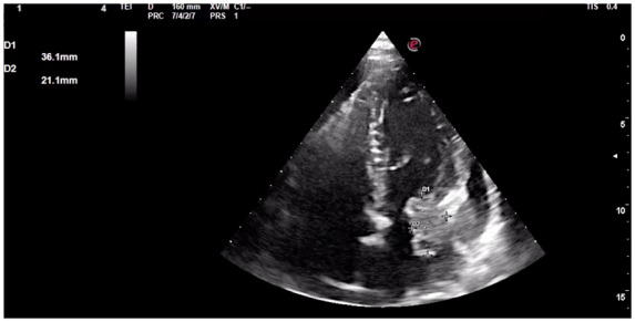
Admission echocardiography showing the apical-four-chamber view of the left ventricle with inhomogeneous left atrial mass (36 mm × 21 mm diameter), adherent to the mitral ring and with irregular borders, suspected for malignancy.
Official websites use .gov
A
.gov website belongs to an official
government organization in the United States.
Secure .gov websites use HTTPS
A lock (
) or https:// means you've safely
connected to the .gov website. Share sensitive
information only on official, secure websites.
Admission echocardiography showing the apical-four-chamber view of the left ventricle with inhomogeneous left atrial mass (36 mm × 21 mm diameter), adherent to the mitral ring and with irregular borders, suspected for malignancy.