Figure 1.

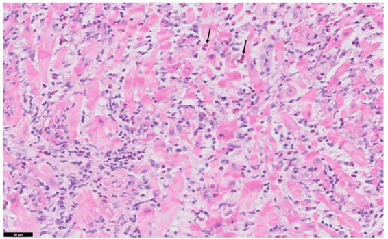
Biopsy from myocardium showing inflammatory cell infiltration with high number of eosinophils (arrow), myocyte destruction, and oedema.

Biopsy from myocardium showing inflammatory cell infiltration with high number of eosinophils (arrow), myocyte destruction, and oedema.