Figure 1.

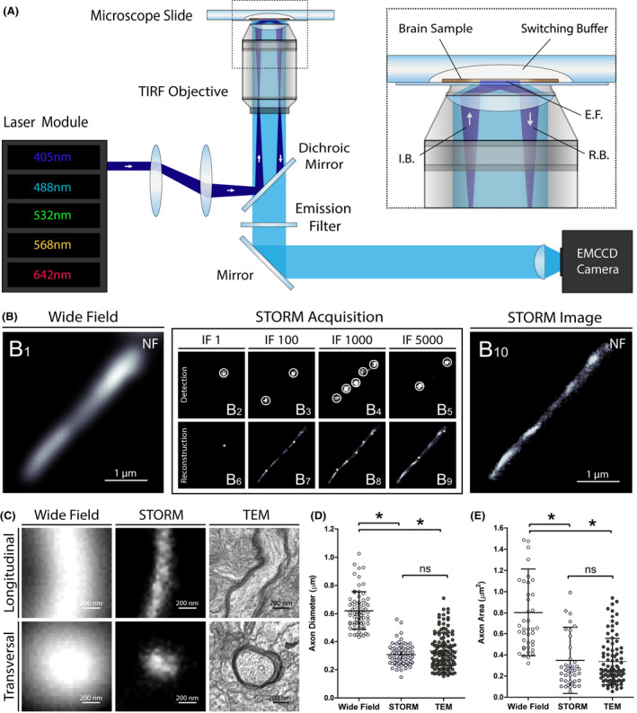
Super‐resolution imaging of human brain samples with STORM. (A) Schematic of the optical setup used for STORM imaging. I.B., incident beam; E.F, evanescent field; R.B., reflected beam. (B) STORM acquisition of a cortical axon in a human brain section immunostained for neurofilaments (NF): a conventional wide field fluorescence microscopy image was first acquired (B1), excitation power was then strongly increased to induce fluorophore blinking and thousands of frames were recorded (B2‐B5). The localization of the activated fluorescent molecules were detected on a per‐frame basis with sub‐pixel accuracy (B6‐B9). The accumulated localizations from all frames were then used to reconstruct a super‐resolution image (B10). IF, imaging frame. (C) Representative images of longitudinally and transversally sectioned prefrontal cortex axons acquired with conventional wide field fluorescence microscopy, STORM and transmission electron microscopy (TEM). (D and E) Axon diameters (longitudinal sections) and areas (transversal sections) measured in human brain using conventional fluorescence microscopy, STORM and TEM. Error bars indicate means with standard deviations. *P < .001
