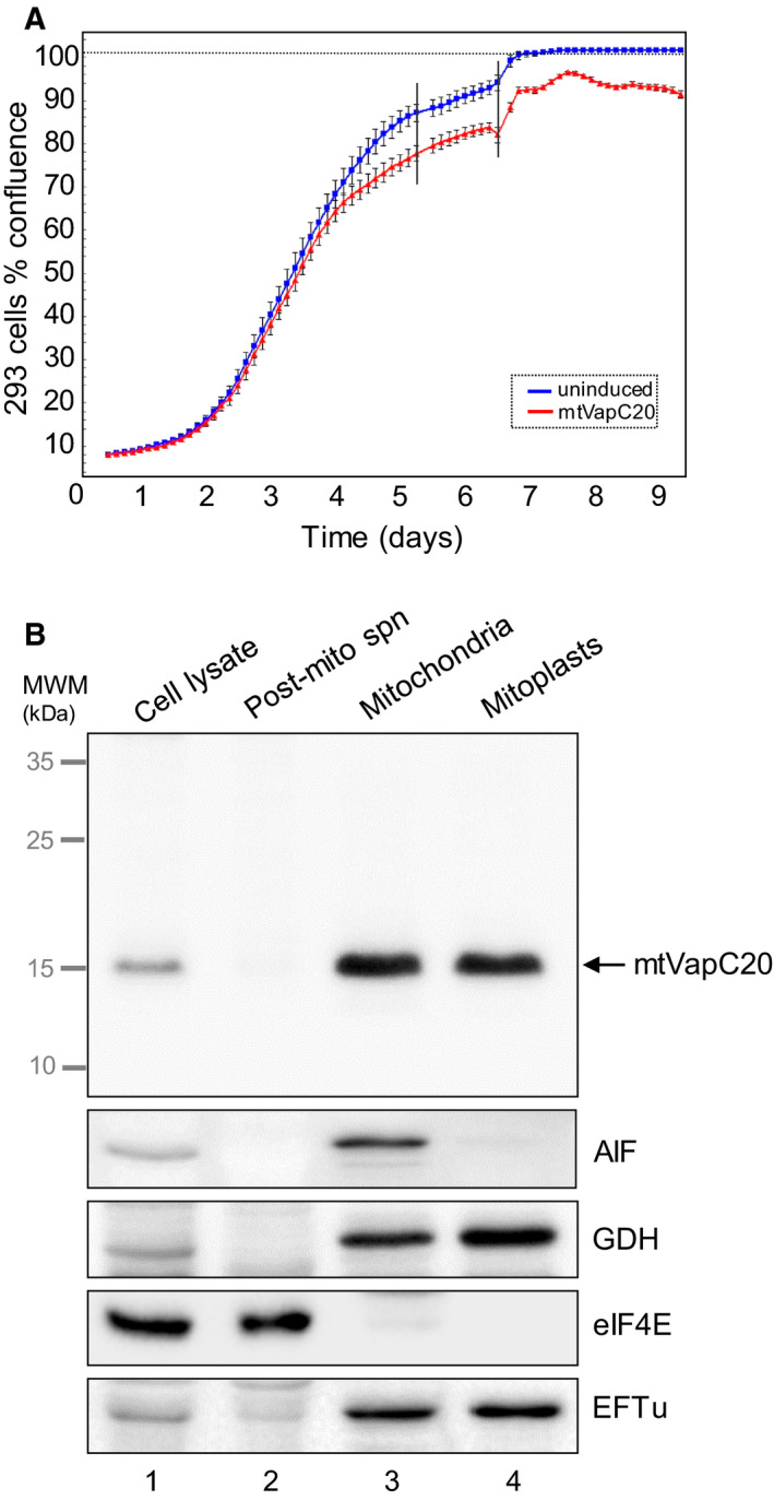
Fig. 1.

mtVapC20 expression and submitochondrial localization. (A) Growth was monitored for 9 days of both uninduced and induced mtVapC20 293 cells (n = 3) using the IncuCyte ZOOM® System. Error bars represent the standard deviation of independent measures (n = 16) carried out every 3 h on each well. Black vertical bars denote the replacement of cell media to prevent acidification. (B) Submitochondrial localization of mtVapC20 was determined by immunoblotting of fractions as described. Anti‐6XHis antibody was used to detect mtVapC20 and the following markers for each mitochondrial compartment: apoptosis‐inducing factor (AIF: mitochondrial intermembrane space), glutamate dehydrogenase and mitochondrial elongation factor Tu (GDH and EFTu: mitochondrial matrix), and eukaryotic initiation factor 4E (eIF4E: cytosol). Data are representative of one experiment. MWM, molecular weight markers.
