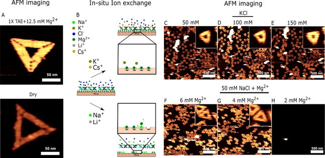
Figure 2.

AFM study of DNA origami adsorption during ionic exchange: A) AFM image of DNA origami in standard folding buffer consisting of 1xTAE (40 mM Tris, 20 mM acetic acid and 1 mM EDTA) and 12.5 mM Mg2+ and in dry after washing with Milli‐Q water. B) Proposed pathway of ionic exchange in mica and the effects on DNA origami adsorption. AFM imaging during in situ buffer exchange with 10 mM Tris, acetic acid, pH 8.0 in addition to 50 (C), 100 (D) and 150 (E) mM KCl and 50/6 (F), 50,4 (G) and 50/2 (H) mM NaCl/Mg2+.
