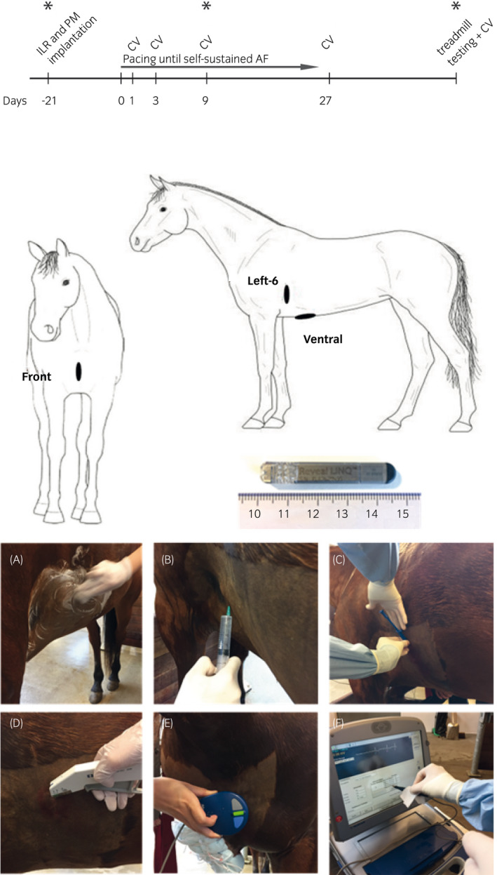
Figure 1.

Schematic illustration of timeline of the study (above), anatomical position of the three implantable loop recorders (middle) and implantation procedure (below). ILR, implantable loop recorder; PM, pacemaker implantation; CV, cardioversion. *Interrogation of the ILR, “Front”: Superficial pectoral region, “Left‐6”: The sixth left intercostal space at the level of the shoulder joint, “Ventral”: The level of the xiphoid process in a cranio‐caudal direction. Picture of the implantable loop recorder used (Medtronic reveal LINQ). Implantation sequence (A‐D) and interrogation with the programmer (E, F) are shown
