
Fig. 1.
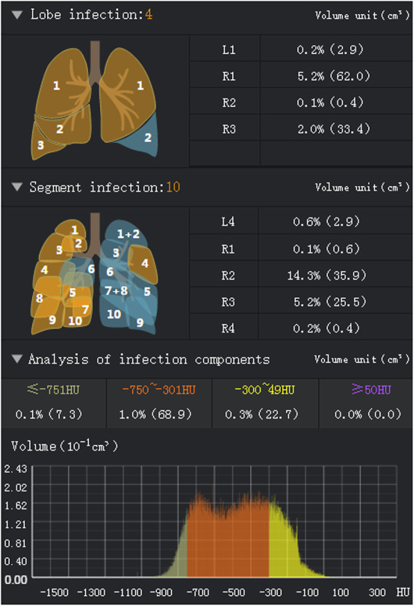
Software interface obtained by inputting original HRCT images of one patient into the DL system. The VOIs and POIs in the lung lobes and bronchopulmonary segments are presented; the HU histogram within the infection regions can be visualized
Official websites use .gov
A
.gov website belongs to an official
government organization in the United States.
Secure .gov websites use HTTPS
A lock (
) or https:// means you've safely
connected to the .gov website. Share sensitive
information only on official, secure websites.
Software interface obtained by inputting original HRCT images of one patient into the DL system. The VOIs and POIs in the lung lobes and bronchopulmonary segments are presented; the HU histogram within the infection regions can be visualized