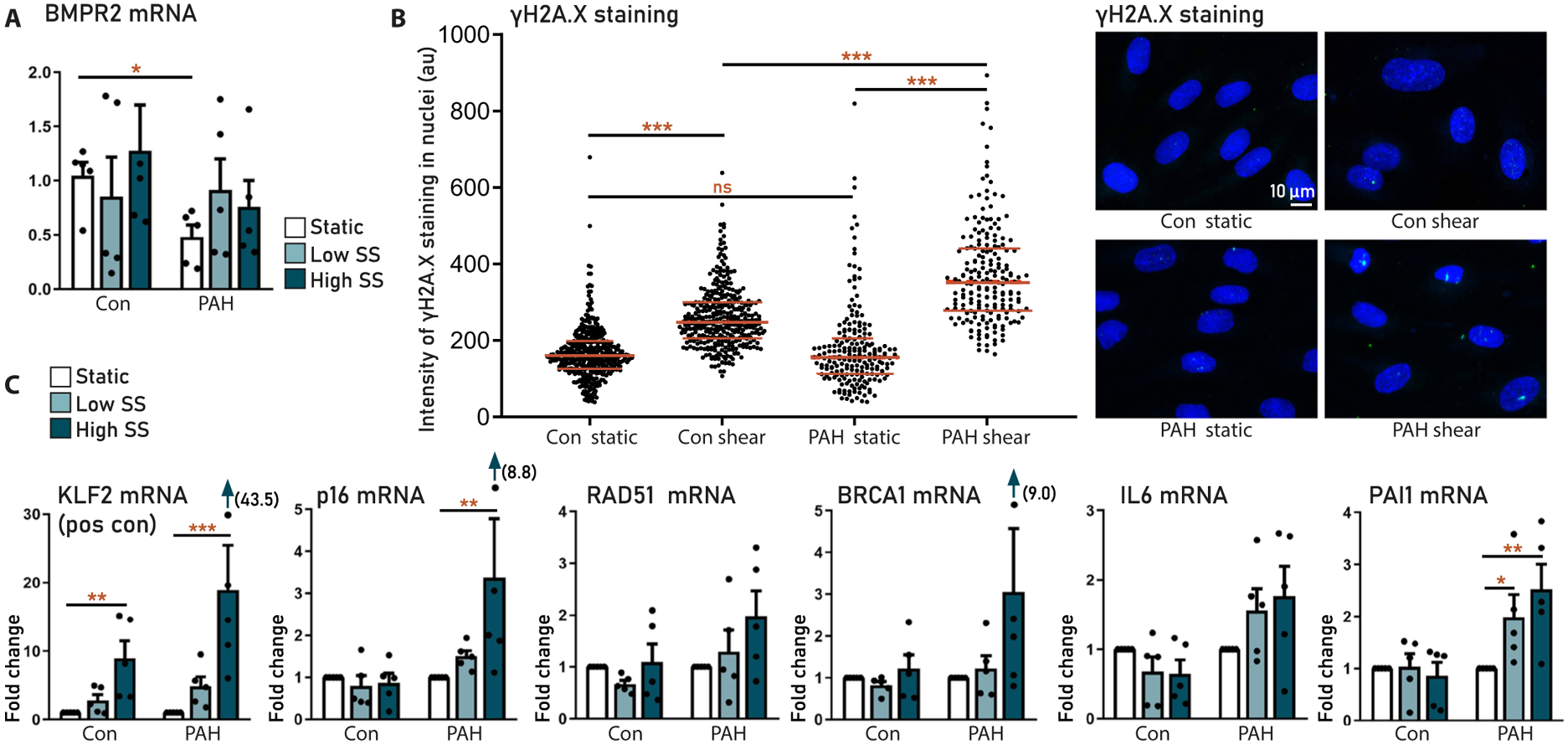
Fig. 6. Prosenescent effects of shear stress on human pulmonary microvascular ECs.

Pulmonary microvascular ECs (pMVECs) isolated from patients with idiopathic PAH (PAH, n = 5) and controls (con, n = 5). (A) BMPR2 expression in control and PAH MVECs in static and low shear stress (2.5 dyne/cm2) and HSS (15 dyne/cm2) conditions. (B) Quantification (left) and representative images (right) of DNA damage (γH2A.X staining) in control and PAH MVECs cultured under static and shear stress conditions. (C) Effect of shear stress on the expression of senescence and DNA damage markers in control and PAH MVECs. Data are reported as means ± SD. Statistics by two-sided t test or Kruskal-Wallis test. Relevant significant differences are indicated with a black bar and asterisk. *P < 0.05, **P < 0.01, ***P < 0.001, and ****P < 0.0001.
