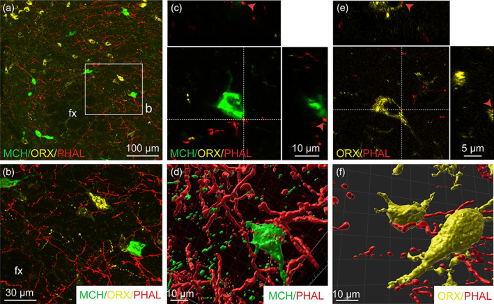
FIGURE 10.

(a) Triple immunofluorescence of PHAL‐labeled axons (red) from the dorsomedial BNST, melanin‐concentrating hormone (MCH, green)‐ and orexin (ORX, yellow)‐positive neurons in the perifornical area and the LHA (experiment BNST#6). (b) High magnification of the inset shown in a. A PHAL‐positive axon (red) is in contact with one MCH‐labeled neuron (c, green) and with one ORX neurons (d, yellow) in the perifornical area. Insets on top in c and d show the orthogonal view on the vertical line and insetson the right show the orthogonal view of the horizontal line. Red arrows indicate the contacting site between PHAL‐positive terminals and MCH or ORX neurons. 3D reconstruction using Imaris software of PHAL‐positive axons (red) from the dorsomedial BNST in contact with MCH (green) and ORX (yellow) neurons in the perifornical area. Fx, fornix. Scale bars are shown in the figure [Color figure can be viewed at wileyonlinelibrary.com]
