FIGURE 13.

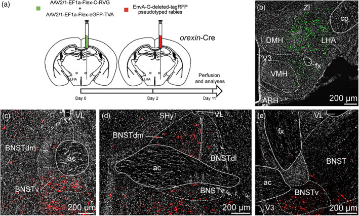
(a) Experimental approach. A mix of AAV‐EF1a‐Flex‐C‐RVG and AAV‐EF1a‐Flex‐eGFP‐TVA was injected at day 0 in the perifornical area and the LHA of 12 weeks old orexin‐Cre male mice. Two days later, mice received injection of EnvA‐G‐deleted‐tagRFP pseudotyped rabies. Animals were perfused 9 days later for further analyses. (b) Photomicrograph of the site of stereotactic injection of the viruses. eGFP‐positive cells (green) were infected with the AAV‐EF1a‐Flex‐eGFP‐TVA. (c–e) Microphotographs illustrating the distribution of neurons projecting onto orexin (ORX) neurons of the perifornical area and the LHA (tRFP, red), at several levels of the anterior bed nucleus of the stria terminalis (BNST). tRFP‐positive cells are observed in the dorsomedial and lateral division of the BNST as wells as in the ventral division. ac, anterior commissure; ARH, arcuate nucleus of the hypothalamus; BNSTdm, dorsomedial division of the BNST; BNSTdl, dorsolateral division of the BNST; BNSTv, ventral division of the BNST; cp, cerebral peduncle; DMH, dorsomedial nucleus of the hypothalamus; fx, fornix; LHA, lateral hypothalamic area; LSv, ventral part of the lateral septum; shy, septohypothalamic nucleus; st, stria terminalis; VMH, ventromedial nucleus of the hypothalamus; VL, lateral ventricle; V3, third ventricle; ZI, zona incerta. Scale bars are shown in the figure [Color figure can be viewed at wileyonlinelibrary.com]
