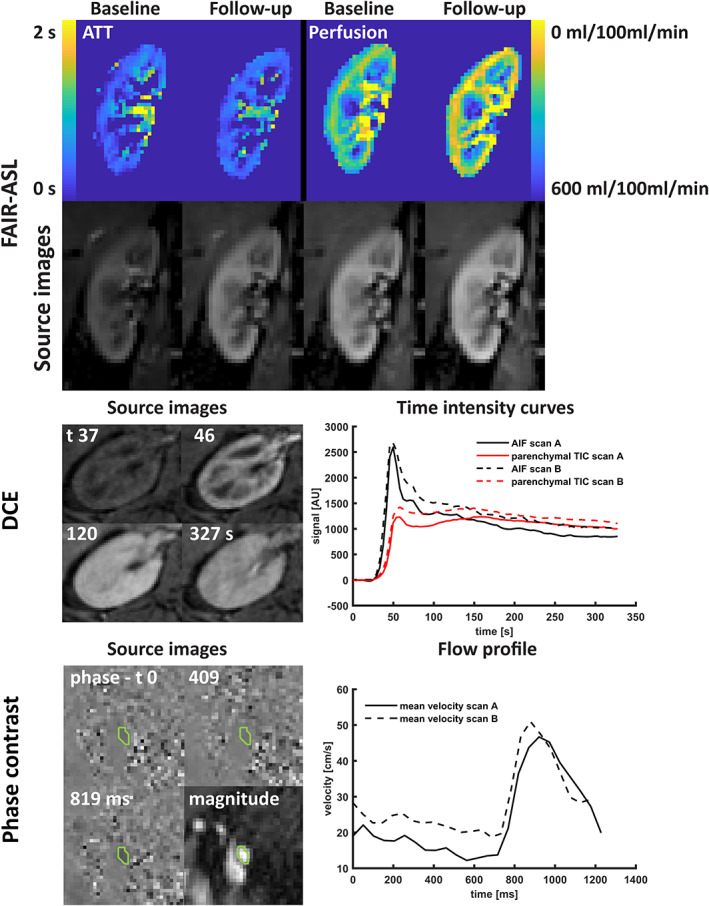
FIGURE 5.

Top: Source images and parameter maps of arterial spin labeling; middle: transverse source images of DCE imaging at four timepoints (precontrast, cortical phase, medullary phase, and late phase) and the AIF and parenchymal TIC measured at both scan sessions; bottom: source images (phase and magnitude) of the phase contrast scan, including the region‐of‐interest and blood flow velocity over the cardiac cycle as measured during the first and second scan session. FAIR‐ASL: flow‐attenuated alternating inversion recovery arterial spin labeling; ATT: arterial transit time; DCE: dynamic contrast enhanced MRI; AIF: arterial input function; TIC: time intensity curve; AU: arbitrary units.
