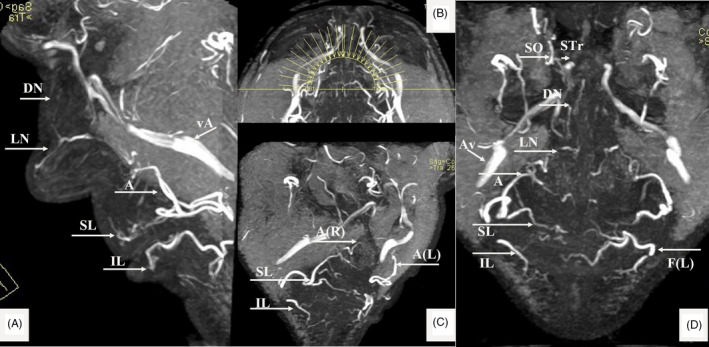
FIGURE 4.

MRA findings (MIP of 3D‐TOF) in a 58‐y‐old male. Superior (SL) and Inferior Labial (IL), Angular (A); Lateral Nasal (LN); Dorsal Nasal (DN); Supratrochlear (STr); Supraorbital (SO) and Facial artery (F); Angular vein (Av); (R) Right and (L) Left. (A) Lateral view. (B) The MIP—reconstruction levels shown on the axial view. (C) Right oblique view with details of the labial arteries. (D) AP view of the (annotated) arteries
