
Fig. 4.
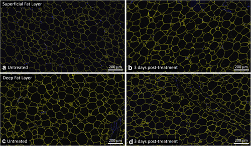
Representative untreated control (a,c) and EMMS‐treated (b,d) tissue from superficial (a,b) and deep (c,d) subcutaneous fat layers, harvested 3 days post‐treatment, subject OKA‐050. Perilipin immunofluorescence stain (TRITC, yellow) and nuclear stain (DAPI, blue), Scale bar = 200 μm. DAPI, 4′,6‐diamidino‐2‐phenylindole; EMMS, electromagnetic muscle stimulation.