
Fig. 9.
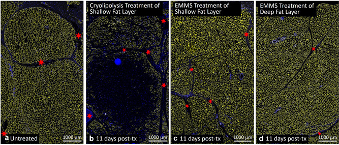
Comparison of (a) untreated control, (b) cryolipolysis, and EMMS‐treated (c) superficial and (d) deep adipose tissue, harvested 11 days post‐treatment. Perilipin immunofluorescence staining (TRITC) shown in yellow. Nuclear stain (DAPI) shown in blue. Scale bar = 1000 μm. Red stars highlight fiber septae or connective tissue structures. DAPI, 4′,6‐diamidino‐2‐phenylindole; EMMS, electromagnetic muscle stimulation.