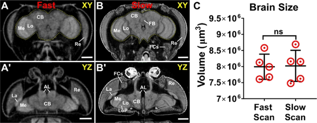
Figure 3: Scanning parameters and image resolution do not alter morphometric analyses.
An adult head scanned using both (A, A’) ‘fast’ scanner settings (hundreds of projections) and (B, B’) ‘slow’ scanner settings (thousands of projections). The brain is outlined in yellow. (C) Brain volume measurements from slow and fast scans. Highlighted structures: AL; antennal lobe; CB, central brain; FB, fan shaped body; FCs, fat cells; La, lamina; Lo, lobula; LoP, lobula plate; Me, medulla; Re, retina. n = 5, Welch’s t-test. ns = not significant. Scale bars = 100 μm. Scanning parameters: Source to Sample Distance (mm): (A) 44.4, (B) 36.5. Source to Camera Distance (mm): (A) 348 (B) 350. Camera Pixel Size (μm): (A-B) 11.6. Image Pixel Size (μm): (A) 2.95, (B) 1.2. This figure has been modified from Schoborg et al.40..

