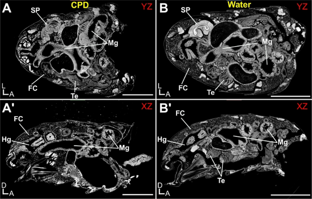
Figure 4: Drosophila melanogaster abdomen imaged by X-ray Microscopy.
Abdomens were stained with 0.5% PTA and imaged hydrated (water) or following critical point drying (CPD). (A) Critical Point Dried abdomen, shown from the YZ perspective and (A’) XZ perspective. (B) Hydrated abdomen, shown from the YZ perspective and (B’) XZ perspective. Various organs are highlighted: FC, fat cells; Hg, hindgut; Mg, Midgut; SP, Sperm Pump; Te, Testes. Scale Bars (A) = 250 μm. D, Dorsal; A, Anterior; L, Left. Scanning parameters: Source to Sample Distance (mm): (A) 6.7, (B) 7. Source to Camera Distance (mm): (A) 28 (B) 29.5. Objective: (A-B) 4X. Image Pixel Size (μm): (A-B) 0.65.

