. 2020 Sep 8;11(37):10265–10278. doi: 10.1039/d0sc02144j

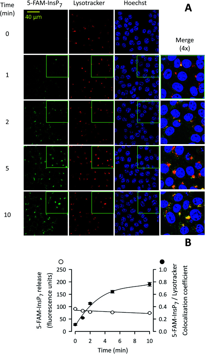
Fig. 7. Analysis by confocal microscopy of the influence of time of LED irradiation of intact cells upon photothermal release of 5-FAM-InsP7. (A) Representative confocal images showing the impact of LED exposure time upon intracellular 5-FAM-InsP7 release. Liposomes (20 μg mL–1) containing 5-FAM-InsP7 were added to cultures of HCT116 cells (0.8 × 10⁶ per well; 1 mL medium; 4 h). Next, cells were washed twice in PBS and irradiated with LED (450 mW; 3 cm) for the indicated times. Cells were then stained with lysotracker and Hoechst. From left to right, the columns of panels show 5-FAM-InsP7 fluorescence (green), lysotracker staining (red), Hoechst staining (blue), and a 4× magnification signal merge; these data are representative images. (B) Mean values and standard deviations for total cellular 5-FAM-InsP7 fluorescence and the degree of lysotracker/5-FAM-InsP7 co-localization (determined at 63× the magnification of the wide-field images), derived from cells treated as described in panel A. Data were derived from 3 biological replicates (each of which involve analyzing 284–340 cells at every time point).

Fig. 7