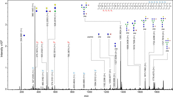
Fig 4. MS/MS spectrum of N-184 glycopeptide from 21K slow prion strain.
The glycopeptide represented in the figure is the one carrying N-glycan with a composition H4N5F2, which was found to be the most abundant structure on this glycosylation site. Fragments of glycan-specific marker ions are represented, together with b and y ions confirming the amino acid sequence of N-184 peptide, and fragments of the peptide backbone with the N-glycan. Blue square–N-acetylglucosamine (GlcNAc), green circle–mannose (Man), red triangle–fucose (Fuc), yellow circle–galactose (Gal), purple diamond–N-acetylneuraminic acid (Neu5Ac).

