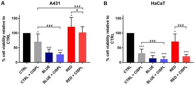
Figure 2.
Analysis of cell viability in (A) A431 and (B) HaCaT cell lines exposed to LED, cisplatinum, and LED and cisplatinum combined. Columns represent mean values and bars represent ± SD (n=3). *P<0.05 and ***P<0.001 vs. CTRL. #P<0.05 and ###P<0.001. LED, light-emitting diode; SD, standard deviation.

